
New NMOSD study points to silent brain changes between relapses
Findings may reshape how doctors monitor stable patients
Written by |
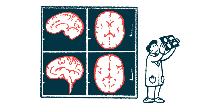




People with neuromyelitis optica spectrum disorder (NMOSD) may experience gradual shrinkage, or atrophy, of the brain even during periods without relapses, according to a new study that challenges the long-held view that most neurological damage in NMOSD occurs mainly during relapses.
The researchers also found that starting biologic therapies earlier and spending a larger share of disease duration on these treatments were associated with slower tissue loss in subcortical gray matter, deep brain regions involved in memory, emotion, and information processing.
Biologic therapies, or those derived from living organisms, currently used in NMOSD include NMOSD-approved therapies and rituximab (sold as Rituxan and others, with biosimilars available).
According to the researchers, these findings could have important implications for how NMOSD is monitored and treated in the future.

Study may shift how NMOSD is monitored
“Our work highlights the need for further studies on cognitive function and for treatment strategies that address not only relapse prevention but also long-term neurodegeneration,” Hiroki Masuda, MD, PhD, the study’s first author and an assistant professor at Chiba University, in Japan, said in a university news release. “Monitoring brain atrophy and cognitive function may become important even in clinically stable patients with NMOSD, as subtle cognitive decline may occur even in the absence of relapses.”
The study, “Biologic therapy and brain atrophy in neuromyelitis optica: a Japanese–German longitudinal MRI study,” was published in the Journal of Neurology, Neurosurgery & Psychiatry.
NMOSD occurs when the immune system mistakenly attacks healthy cells in the brain and spinal cord. In most cases, the disease is associated with self-reactive antibodies targeting AQP4, a protein mainly found in neuron-supporting cells.
These autoimmune attacks trigger episodes of inflammation and nerve damage, known as relapses, that can cause new or worsening NMOSD symptoms. Over time, relapses can lead to permanent disability.
For a long time, NMOSD has been viewed as a disease in which most neurological damage occurs during relapses. However, growing evidence suggests that brain and spinal cord tissue may continue to shrink even when patients remain relapse-free.
Although biologic therapies are highly effective at preventing relapses, it has remained unclear whether they also protect against this ongoing tissue loss.
MRI scans tracked brain changes over time
To answer these questions, a team led by researchers at Chiba University set out to compare changes in brain volume between 72 adults with AQP4-related NMOSD who had no relapses between two MRI scans and 52 age- and sex-matched healthy volunteers.
They also investigated whether brain tissue loss was linked to disease characteristics and whether biologic therapies were associated with changes in brain atrophy.
The patient group was composed of 63 people treated at Chiba University Hospital and nine enrolled through the Berlin Registry of Neuroimmunological Entities, an ongoing research registry that collects clinical data from people with immune-mediated neurological diseases.
Most patients were women (84.7%), had a median age of 53.5 years, and had been living with NMOSD for a median of about eight years. Both NMOSD patients and healthy volunteers underwent two brain MRI scans a median of 1.2 and 1.4 years apart, respectively.
At the first MRI scan, participants with NMOSD had significantly lower volumes of cortical gray matter, which makes up the brain’s outer layer, and lower total gray matter than healthy volunteers.
Within the NMOSD group, smaller volumes of subcortical gray matter — deep brain structures involved in memory, emotion, and information processing — were significantly associated with higher disability scores, a higher number of previous relapses, and an older age at disease onset.
By the follow-up scan, NMOSD patients also had significantly lower overall brain volume than healthy volunteers. Further analysis showed that, during follow-up, people with NMOSD lost brain tissue significantly faster than healthy volunteers despite remaining relapse-free.
Brain tissue loss occurred despite no relapses
Specifically, whole-brain volume shrank by a median of 0.44% per year in people with NMOSD, compared with an essentially stable rate of -0.19% per year in healthy volunteers. Faster tissue loss also occurred in the parietal and temporal lobes, brain regions involved in sensory processing, language, and memory.
“These findings suggest that neurodegeneration in NMOSD may occur through mechanisms independent of clinical relapses,” Masuda said. “However, the association between subcortical gray matter volume and neurological disability should be interpreted carefully, because subcortical gray matter volume was also associated with disease duration and relapse number, which may partly reflect cumulative disease burden over time.”
Overall brain atrophy rates were similar between patients who received biologic therapy throughout the study and those who received other treatments. However, spending a larger share of disease duration on biologic therapy was significantly linked to slower subcortical gray matter atrophy. Likewise, earlier initiation of biologic treatment was associated with lower rates of subcortical gray matter loss over time.
“This study suggested silent brain atrophy in patients with [AQP4-related NMOSD],” the researchers wrote. They added that earlier and sustained use of biologic therapies may be linked to slower loss in subcortical gray matter, “highlighting the need to consider long-term treatment strategies and to further investigate factors that contribute to brain atrophy.”




Leave a comment
Fill in the required fields to post. Your email address will not be published.