NMOSD patients face spinal cord shrinkage even absent relapses
Study: Rituximab may slow atrophy better than other immunosuppressives
Written by |
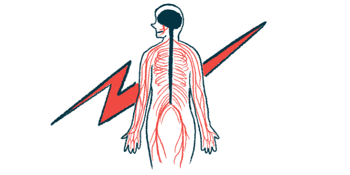


Even if they don’t have disease relapses, people with neuromyelitis optica spectrum disorder (NMOSD) tend to experience abnormal atrophy, or shrinkage, of the spinal cord over time, a new study shows.
According to the researchers, “spinal cord involvement … accounts for significant disability in up to 80% of patients with NMOSD,” making ways to slow atrophy important for people with the rare autoimmune disorder.
The new findings also suggest that the immunosuppressive therapy rituximab may work better than other similar drugs to slow spinal cord shrinkage, the scientists noted.
Still, the team cautioned that the study was limited to only a few dozen patients, so further work is needed to validate the results.
The study, “Longitudinal Spinal Cord Atrophy in Patients With Neuromyelitis Optica Spectrum Disorder and Its Association With Rituximab Treatment,” was published in the journal Neurology.

In NMOSD, the immune system attacks healthy cells in the brain and spinal cord. Most cases are associated with self-reactive antibodies that target AQP4, a protein mainly found in neuron-supporting cells.
The disease is marked by relapses, or periods when NMOSD symptoms suddenly worsen or return, interspersed with periods of remission, during which symptoms are reduced or absent.
Little data on spinal cord shrinkage over time in NMOSD
Previous research has suggested that NMOSD is marked by spinal cord atrophy, but few studies have tracked how this type of shrinkage progresses over time in people with the condition.
To learn more, a team of scientists in China analyzed 233 MRI scans taken over a median follow-up time of nearly two years in 72 people with NMOSD. Most of these patients (85%) were female and tested positive (75%) for anti-AQP4 antibodies. The patients’ mean age was 40, and they had been living with NMOSD for a median of slightly longer than 1.5 years.
The researchers noted that all of the patients were in clinical remission and experienced no relapses or treatment changes during the study period. A total of 63% had MRI follow-up for more than two years.
Three measures of spinal cord volume were analyzed: cross-sectional area, or CSA, which looks at the total volume in two-dimensional slices of the spinal cord; anteroposterior (AP), or front-to-back measurements; and right-left, or RL, measurements.
The measures from the NMOSD patients were compared against MRI reference values derived from scans from more than 2,000 healthy people. The results showed that, at initial MRI scans, all three spinal cord measures were comparable between the groups.
Over time, however, those with NMOSD showed significantly more spinal cord atrophy per year than healthy people across all three measures. Specifically, by 0.27 for CSA, 0.23 for AP, and 0.14 for RL.
This longitudinal study demonstrates that spinal cord atrophy in NMOSD progresses even in the absence of clinical relapses.
According to the team, the patients experienced estimated annual percentage volume reduction rates over time of 2.54% for CSA, 1.78% for AP, and 0.77% for RL, compared with the first MRI.
“This longitudinal study demonstrates that spinal cord atrophy in NMOSD progresses even in the absence of clinical relapses,” the researchers wrote.
Subgroup analyses showed that spinal cord atrophy relative to reference values was observed only in patients with spinal cord inflammation, called transverse myelitis, and those testing positive for anti-AQP4 antibodies.
The team also found that higher blood levels of neurofilament light chain (NfL) and glial fibrillary acidic protein (GFAP), two markers of brain cell damage, were significantly associated with faster spinal cord atrophy.
“Elevated [blood] GFAP and NfL levels, especially during follow-up, were associated with faster spinal cord atrophy rates, highlighting their potential as biomarkers for ongoing spinal cord atrophy in NMOSD,” the researchers wrote.
Statistical analyses adjusted for potential influencing factors showed that a prolonged interval between initial transverse myelitis and initial MRI was an independent predictor of slower CSA atrophy. In addition, older age and longer lesion length independently predicted faster RL atrophy. No independent predictors of AP atrophy rate were identified.

Atrophy was slower for patients on rituximab vs. other treatments
Nearly 70% of the NMOSD patients were on rituximab, an antibody-based therapy marketed as Rituxan in the U.S. and Mabthera in Europe (with biosimilars available).
Rituximab works by killing B-cells, the immune cells that produce antibodies, including those that drive NMOSD. Although the therapy is not approved as an NMOSD treatment, it is commonly used off-label to help manage the disease.
The remaining patients, nearly one-third in all, were taking other immunosuppressive therapies, namely mycophenolate mofetil, azathioprine, or prednisone.
Statistical analyses demonstrated that rituximab-treated patients had significantly slower RL atrophy than those on other therapies. CSA showed a similar trend, though the difference didn’t reach statistical significance, meaning that it could be due to random chance. Rates of AP atrophy were comparable between patients on rituximab and those on other drugs.
“Although further validation is needed, our findings provide preliminary support for a possible structural benefit of rituximab beyond relapse prevention, particularly in preserving … spinal cord architecture,” the researchers wrote, noting a need for further studies to see if rituximab can better preserve the spinal cord in NMOSD.
Overall, the findings “advance our understanding of NMOSD [underlying mechanisms] and underscore the potential of integrating [blood] and imaging biomarkers to enhance individualized monitoring and guide early, targeted therapeutic strategies,” the team concluded.