
NMOSD patients report lower sleep quality, study shows
They also spend more time in bed and sleeping than healthy people
Written by |
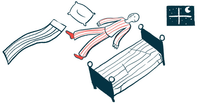




People with neuromyelitis optica spectrum disorder (NMOSD) report a greater burden of sleep problems and, when objectively monitored, spend more time in bed and sleeping than healthy people, according to a small study from Thailand.
Data from 24 adults with NMOSD and self-reactive antibodies against the aquaporin-4 (AQP4) protein — the most common type of NMOSD-driving antibody — also showed that a higher body mass index (BMI), a proxy measure of body fat based on height and weight, was associated with poorer sleep efficiency and taking longer to fall asleep.
Brain MRI scans also showed reduced volume in certain brain regions, but the study found no clear link between these volume differences and sleep changes.
“These hypothesis-generating findings are consistent with a multifactorial pattern of associations and highlight the need for longitudinal studies integrating objective sleep measurement, neuroimaging and biomarkers.” researchers wrote.
The study, “Multimodal assessment of sleep and regional brain volumes in AQP4-IgG-positive neuromyelitis optica spectrum disorder: an exploratory cross-sectional study,” was published in the journal Multiple Sclerosis and Related Disorders.
Most NMOSD patients experience sleep disturbances
NMOSD is a rare autoimmune disease characterized by damaging inflammation, primarily in the spinal cord and optic nerves, which relay signals between the eyes and the brain, although the brain can also be involved.
In most cases, the immune system wrongly produces self-reactive antibodies against AQP4, a protein found at high levels on nerve-supporting cells called astrocytes. Given that AQP4 is found in brain regions involved in sleep, including the hypothalamus and thalamus, inflammatory attacks may negatively influence sleep in people with NMOSD.
Evidence suggests that most people living with NMOSD experience sleep disturbances. However, studies combining patients’ own reports with objective sleep monitoring and detailed brain imaging have been limited.
With this in mind, a team of clinicians in Thailand analyzed subjective and objective reports of sleep and wake patterns from 24 adults with clinically stable NMOSD and anti-AQP4 antibodies. They also aimed to determine whether sleep measures were associated with changes in the volume of specific brain regions.
Higher body mass index associated with lower sleep efficiency
Participants had not experienced a disease relapse in the previous six months. Sleep data from 26 healthy people were also analyzed as controls. Because patients were significantly older than healthy controls (mean 49.7 vs. 33 years), analyses of key sleep measures were adjusted for age as well as BMI.
Participants completed questionnaires assessing sleep quality, daytime sleepiness, and quality of life, and wore a wrist-worn device for seven consecutive days that recorded rest-activity cycles.
Wrist-worn device data were used to estimate measures such as total sleep time, time spent in bed, time to fall asleep, sleep efficiency — the proportion of time in bed actually spent asleep — and nighttime awakenings.
Statistical analyses adjusted for age and BMI showed that people with NMOSD spent significantly more time in bed, by 57.8 minutes, and slept significantly more, by 50 minutes, than controls.
Overall sleep efficiency did not significantly differ between the groups. Patients also reported significantly poorer pain-related quality of life, as well as more sleep disturbances, but this difference failed to reach statistical significance.
Within the NMOSD group, additional analyses indicated that higher BMI was significantly associated with lower sleep efficiency and longer time to fall asleep. This association between higher BMI and poorer sleep efficiency remained after accounting for BMI and disability, as measured by the Expanded Disability Status Scale.
These findings support a multifactorial pattern of sleep disturbance involving behavioural, clinical, and neurobiological factors.
The clinicians also compared brain MRI scans from 20 of the NMOSD patients with those from 20 age-matched healthy people drawn from an institutional database. The scans were processed to measure the volumes of 256 brain regions, including structures involved in sleep regulation.
Corrected analyses demonstrated that NMOSD patients had significantly smaller volumes in the hippocampus, a brain region important for memory, and corpus callosum, the bundle of nerve fibers connecting the brain’s two hemispheres. Certain regions of the thalamus also showed possible reductions, although these findings did not meet the study’s stricter threshold for significance.
When the researchers tested whether the volume of individual brain regions was related to sleep parameters, several apparent associations emerged. But none remained significant after standard correction methods, meaning the study did not establish that the structural brain differences were responsible for the sleep changes.
The findings highlight that, “across modalities, sleep disruption was characterized by greater subjective sleep burden and altered sleep-wake timing, accompanied by selective structural brain differences,” the team wrote. “These findings support a multifactorial pattern of sleep disturbance involving behavioural, clinical, and neurobiological factors.”
Among the study limitations were the small sample size and its cross-sectional design, which looks at data at a single point in time, as well as the absence of overnight polysomnography — the gold standard test for assessing sleep.
“Future longitudinal studies with … matched cohorts, concurrent polysomnography, and integrated biomarker assessment will be required to better characterize the temporal and mechanistic relationships among brain structure, sleep disruption, and disease activity,” the researchers wrote.




Chuck Visnic
Sleep and night sweats are common good sleeping is in the past before NMOSD.
I think memories are more active replaces dreams.