Rituximab Clears NMOSD-causing Antibodies From Lymph Nodes
Written by |


Self-reactive antibodies that cause most cases of neuromyelitis optica spectrum disorder (NMOSD) are released directly from immune B-cells in lymph nodes as opposed to cells in the bloodstream, a study has discovered.
These findings implicate ongoing lymph node activity as the main driver of disease-causing antibody production in NMOSD.
Moreover, treatment with rituximab, a therapy used off-label by some NMOSD patients to prevent relapses, effectively eliminated self-reactive B-cells and the antibodies they produced directly from lymph nodes, leaving the bloodstream antibodies untouched.
These data may explain rituximab’s effectiveness in several antibody-related diseases and highlight the potential value of direct lymph node measurements across autoimmune conditions such as NMOSD, the researchers noted.
The study, “Rituximab abrogates aquaporin-4–specific germinal center activity in patients with neuromyelitis optica spectrum disorders,” was published in the Proceedings of the National Academy of Sciences.

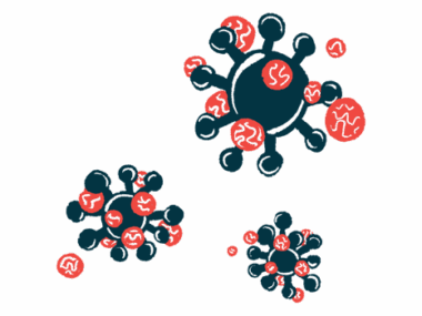
In about 70% of NMOSD cases, self-reactive antibodies, or autoantibodies, target and damage aquaporin-4 (AQP4), a water channel protein commonly found in the brain, spinal cord, and optic nerve.
People with NMOSD have periodic episodes (relapses) of immune activation and autoantibody production, triggering inflammation of the optic nerve and causing pain, blurry vision, or vision loss, as well as the spinal cord, which can lead to a wide range of symptoms.
Rituximab (sold as Rituxan among others) is commonly used off-label in NMOSD. Administered by an into-the-vein infusion, it prevents or reduces relapses by depleting antibody-producing B-cells that carry the protein CD20.
In NMOSD patients treated with rituximab, the levels of anti-AQP4 antibodies in their bloodstream remain unaffected, which seems unusual given the therapy’s effectiveness.
Antibody-producing B-cells start in small areas within lymph nodes called germinal centers, where, when activated, they divide and mature to generate high-affinity antibodies. After reaching maturity, B-cells exit the lymph nodes and enter the bloodstream to become plasma B-cells, providing long-term antibody production and immunity against infection (or autoantibodies and autoimmune disease).
Alternatively, some studies suggest ongoing activity within lymph node germinal centers, not plasma B-cells, may be the source of some autoantibodies, including those in NMOSD patients. Furthermore, the role of germinal centers in NMOSD has not been explored directly.
Researchers at the University of Oxford, U.K. investigated whether AQP4-autoantibody production in germinal centers is a core feature of NMOSD that could be stopped by rituximab. The research team recruited 63 people with NMOSDs, of whom 35 had been treated with rituximab.
Treatment led to a significant reduction in annual relapse rates (ARR), with 31 of the 35 (89%) stopping other immunotherapies. The levels of AQP4-autoantibodies in their bloodstream remained unchanged with rituximab.
Blood samples were collected to measure levels of two types of anti-AQP4 autoantibodies — immunoglobulin M (IgM) and subclasses of immunoglobulin G (IgG) — whose presence is associated with germinal center activity. The team then compared these markers to episodes of symptom relapse.
The presence of either AQP4-IgM or a shift from the AQP4-IgG1 subclass was associated with a sixfold increased risk of relapse, which “implicate [germinal center] activity as a source of AQP4 antibody production around the time of relapses,” the research team wrote.
Fluid was then collected (aspirated) from the deep cervical lymph nodes (dCLNs) in the neck, which, as part of the local lymphatic system, receive drained fluids from the brain and spinal cord. In total, 36 dCLN aspirations were performed in 14 NMOSD patients and 14 controls.
As anticipated, no anti-AQP4 autoantibodies were found in blood or dCLN aspirates from the 14 controls.
However, in all seven NMOSD patients who were not given rituximab, anti-AQP4 IgG autoantibodies were found in their dCLN aspirates. Compared to total IgG levels in the lymph nodes, these anti-AQP4 autoantibodies were about 200 times higher than in blood, “suggesting local (“intranodal”) synthesis of AQP4-IgG,” the researchers said.
In comparison, dCLN aspirate samples collected at varied times after rituximab treatment found anti-AQP4 autoantibodies in two of 11 (18%) NMOSD patients.
These results showed “intranodal synthesis of AQP4-IgG is effectively abrogated by [rituximab] administration,” the researchers wrote. By extension, they suggest “AQP4-specific B cells exist within NMOSD patient dCLNs before [rituximab] administration.”
The depletion of B-cells within lymph nodes persisted for several months after rituximab, mainly in those who received more than one infusion compared to patients who were given a single infusion or no treatment.
To validate these results over time within individuals, AQP4-autoantibodies and B-cell levels were assessed from four patients who underwent blood and dCLN sampling.
In all cases, three to 12 months after rituximab, AQP4-autoantibodies remained unchanged in the bloodstream, whereas in the dCLNs, both B-cells and autoantibodies fell to undetectable levels. “Hence, [rituximab] robustly depleted B cells and AQP4-IgGs in dCLNs without changes in [blood] AQP4-IgGs,” the researchers said.
“Our findings implicate ongoing [germinal center] activity as a rituximab-sensitive driver of AQP4 antibody production,” they said. “They may explain rituximab’s clinical efficacy in several autoantibody-mediated diseases and highlight the potential value of direct [germinal center] measurements across autoimmune conditions.”