Optic Nerve Fiber Damage After Acute NMOSD Attacks Studied
Damage is greater in NMOSD than in idiopathic optic neuritis, study shows
Written by |

Optic nerve fiber damage is usually more severe in people with neuromyelitis optica spectrum disorder (NMOSD) than with idiopathic optic neuritis, based on a clinical measure called visual evoked potential, a study shows.
Results also suggest the recovery window after an acute NMOSD attack is shorter, highlighting the importance of prompt treatment.
The study, “Different visual evoked potentials in neuromyelitis optica spectrum disorder-related optic neuritis and idiopathic demyelinating optic neuritis: a prospective longitudinal analysis,” was published in BMC Ophthalmology.
NMOSD is often marked by optic neuritis wherein the optic nerves, which relay signals from light-sensing cells in the eyes to visual processing centers in the brain, become inflamed. NMOSD-associated optic neuritis, referred to as NMOSD-ON, can cause eye pain and loss of visual acuity.
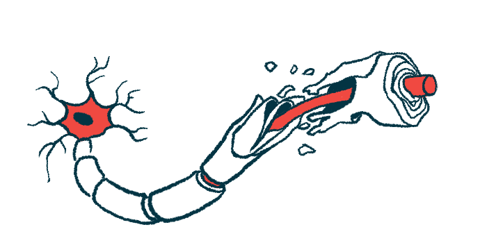
Visual evoked potential, or VEP, measures how fast electrical signals can move in response to visual stimulation. In patients with optic neuritis, VEP can indicate nerve fiber damage and loss of myelin, the protective sheath that insulates nerve fibers.
Scientists at Sun Yat-Sen University, China researched how VEP is affected in NMOSD-ON. VEP was measured in a standard setup where patients looked at a black-and-white checkerboard of two sizes, 15 and 60 inches.
“Little is known about VEP changes in NMOSD-ON patients with an acute attack … With the development of novel NMOSD-ON treatments, it is necessary to describe the natural VEP pattern in NMOSD-ON using different check sizes,” the researchers wrote.
The study included 26 people with NMOSD who were within 30 days of an acute attack causing optic neuritis. All were positive for antibodies against AQP4. The analysis also included 58 people with optic neuritis of unexplained origin, or idiopathic optic neuritis (IDON).
All received treatment with high-dose methylprednisolone followed by prednisone, which was tapered off after at least six months. Both prednisone and azathioprine were prescribed for maintenance treatment.
Compared to IDON patients, a significantly higher proportion of those with NMOSD-ON were female (84.6% vs. 55.2%) and had experienced previous episodes of optic neuritis (34.6% vs. 6.9%). Other characteristics were generally comparable. The mean age in both groups was early 30s.
Participants were assessed for VEP at the study’s start, then again after one, three, and six months. Visual acuity was also tested each time.
There was no significant difference in visual acuity scores between the groups at any time. In both groups, visual acuity generally got better immediately after an acute attack, though the researchers noted the recovery periods were typically shorter in NMOSD-ON compared to IDON with significant improvements only seen in the first month among NMOSD-ON patients.
“Such a short recovery window indicated that more prompt treatment in NMOSD-ON is needed,” they wrote.
In terms of overall VEP patterns, significantly more patients with IDON than NMOSD-ON had normal patterns after three months (24% vs. 4.5%). In both groups, the most common VEP abnormality was delayed latency, meaning it took unusually long for electric signals to travel along the optic nerves from the eyes to the brain.
In a VEP-based measure called P100 amplitude, NMOSD-ON patients showed no significant improvement during follow-up, but IDON patients showed improvements at one and three months. At six months, average P100 amplitude was significantly lower in NMOSD-ON patients than IDON patients.
These between-group differences in P100 amplitude were only seen in assessments using the 15-inch checkerboard. P100 amplitude measures using the 60-inch check showed no significant difference between the groups at any point.
“Our results demonstrated that the NMOSD-ON patients have more severe [nerve fiber] damage than the IDON patients regarding the P100 amplitude and abnormal VEP response pattern,” the researchers said, noting that “a small check size was more sensitive to detecting abnormality in NMOSD-ON than a large check size.”
P100 amplitude measures correlated significantly with measures of visual acuity among the NMOSD-ON patients, statistical analyses showed.
The small sample size and short follow-up were mentioned as the study’s limitations.


